Digital impressions
Photogrammetry vs. Intraoral Scanning for Full-Arch Implant Cases
Compare complete-arch implant capture methods, understand the evidence limits and prepare a clear digital handoff for your dental laboratory.
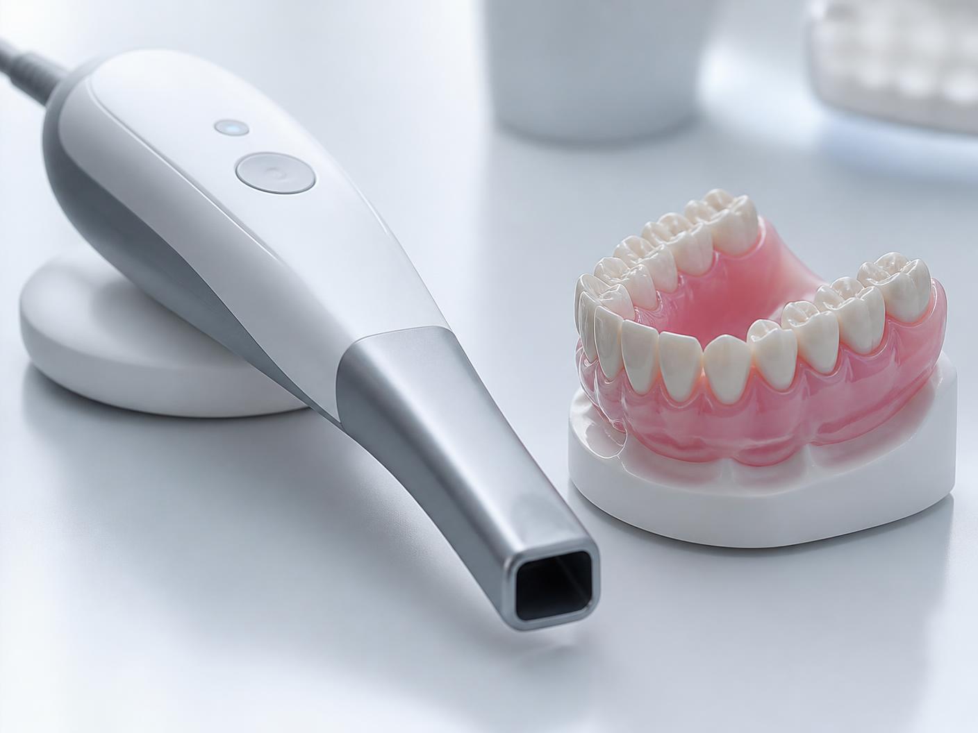
Photogrammetry and intraoral scanning capture different information within a complete-arch workflow. Select the method and verification protocol for the specific case, equipment and restorative plan; neither method guarantees accurate seating in every clinical situation.
What is the difference between the two methods?
Intraoral scanning builds a digital surface record from images acquired inside the mouth. Scan bodies help represent implant relationships within an implant-restorative workflow. Complete-arch capture requires attention to the selected system, scan protocol and the information needed by the laboratory.
Dental photogrammetry uses identifiable targets to record implant relationships. Implant-coordinate capture is not the same as capturing the surrounding tissue surfaces or all restorative records. A case may therefore use photogrammetry together with intraoral scans rather than treating the methods as mutually exclusive.
What does the research say—and what does it not say?
A 2025 systematic review and meta-analysis compared photogrammetry and intraoral scanning in complete-arch digital implant impressions. The review included 13 studies, only three of which were in vivo, and reported methodological heterogeneity. Its findings should be interpreted in the context of the evaluated equipment and study methods.
A separate 2021 review of full-arch digital implant impressions included 30 studies, of which 29 were in vitro. The predominance of laboratory studies limits the conclusions that can be drawn about routine clinical outcomes. These publications are useful for a case-specific discussion, not for promising that a device will eliminate remakes or deliver passive fit in every patient.
Agree on the capture protocol before the appointment
Tell the laboratory which scanner or photogrammetry system you use, which restorative components are planned and how the files will be exported. Ask the coordinator to identify any additional tissue or restorative records needed. Confirm that the intended files are readable and distinguish missing information from a complete submission.
Megatec states that it accepts iTero and TRIOS files and integrates iCam4D, PIC and other photogrammetry data. For its permanent-restoration workflow, a tissue scan is required. Contact the lab for the specific handoff rather than assuming an implant-position file includes every required surface.
- Identify the arch, implant sites and restorative components.
- Confirm system compatibility and export format with the lab.
- Include the required tissue scan and case prescription.
- Flag incomplete, uncertain or revised capture data.
- Confirm receipt before the case moves into design.
Digital capture does not replace clinical verification
An attractive digital model is not proof of a clinically acceptable final restoration. Verification and seating decisions remain with the treating clinician. If records or seating raise a concern, coordinate with the lab before proceeding rather than assuming either the capture method or the prosthesis is responsible.
This article is a workflow overview for dental professionals, not a device-specific scanning protocol or patient treatment recommendation. Use the system manufacturer’s instructions and your clinical judgment, and resolve case-specific questions with the laboratory.
Sources & further reading
- Photogrammetry Versus Intraoral Scanning in Complete-Arch Digital Implant Impression: A Systematic Review and Meta-Analysis
2025 review; mixed study designs and methodological heterogeneity limit universal conclusions.
- Accuracy of full-arch digital implant impressions taken using intraoral scanners and related variables: A systematic review
2021 review; evidence consisted predominantly of in-vitro studies.
Educational content from Megatec Dental Laboratory. Company service descriptions are not independent clinical evidence. Individual treatment, verification and maintenance decisions remain with the treating clinician.